There are no items in your cart.
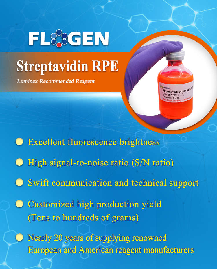
Streptavidin RPE
Numbering
Product Overview
Biotinylated nucleic acids or antibodies can be effectively detected using streptavidin-fluorophore conjugates. Streptavidin (SA) is a tetrameric protein capable of binding up to four biotin molecules with exceptionally high affinity. Phycoerythrin (PE), a fluorescent protein derived from red algae, exhibits strong fluorescence with absorption peaks at 496 nm, 546 nm, and 565 nm, and a maximum emission at 578 nm.
Streptavidin-RPE (SAPE) combines the high biotin-binding capacity of streptavidin with the bright fluorescence of PE, making it an ideal reagent for biotin-streptavidin detection systems. It is widely used in:
-
Flow cytometry
-
Bead-based multiplex assays (e.g., Luminex xMAP®, Illumina VeraCode®)
-
Microarray platforms (e.g., Affymetrix GeneChip®)
-
Immunohistochemistry (IHC)
-
Microplate-based immunoassays
This conjugate delivers high sensitivity and reliable performance across a variety of fluorescence-based detection applications.
Features and Advantages
Consistent Lot-to-Lot Quality
Self Made Fresh and High Quality Materials for Conjugation
Strict QC Criteria
Biotin-Binding and Fluorescent Intensity Check
Flexible Production Capacity
Well-Experienced Team for Conjugation
Unique Process for Fast and Large-Scale Purification
Application
For Flow Cytometry
Streptavidin-based amplification techniques are widely used in flow cytometry for increased signal output and greater sensitivity. Phycobiliprotein-conjugated streptavidin such as SA-PE, and SA-clAPC are used to detect biotinylated biomolecules such as primary and secondary antibodies.
For Microarray Applications
In microarray assays, arrays of oligonucleotides are immobilized onto gene chips to capture target sequences. Gene expression is detected by labeling complementary RNA (cRNA) with biotin, followed by hybridization with the immobilized probes. After hybridization, the array is stained with Streptavidin-RPE (SAPE), enabling detection through the strong fluorescence signal of phycoerythrin (PE). The resulting fluorescent intensity is then quantified and analyzed using specialized software to determine gene expression profiles.
Application
For Flow Cytometry
Streptavidin-based amplification techniques are widely used in flow cytometry for increased signal output and greater sensitivity. Phycobiliprotein-conjugated streptavidin such as SA-PE, and SA-clAPC are used to detect biotinylated biomolecules such as primary and secondary antibodies.
| Label | Excitation | Emission |
| R-Phycoerytherin (RPE) | 496, 546, 565 | 578 |
| Crosslinked APC (cl-APC) | 650 | 660 |
| FITC | 490 | 525 |
For Microarray Applications
In microarray assays, arrays of oligonucleotides are immobilized onto gene chips to capture target sequences. Gene expression is detected by labeling complementary RNA (cRNA) with biotin, followed by hybridization with the immobilized probes. After hybridization, the array is stained with Streptavidin-RPE (SAPE), enabling detection through the strong fluorescence signal of phycoerythrin (PE). The resulting fluorescent intensity is then quantified and analyzed using specialized software to determine gene expression profiles.
For Bead or Microsphere-Based Assays (e.g., Luminex xMAP®, Illumina® VeraCode®)
Beads functionalized with carboxyl groups or amine linkers are coupled to antibodies or nucleic acids for target capture. The target protein or complementary oligonucleotide is biotinylated and subsequently binds to the capture molecule immobilized on the bead surface through agglutination or hybridization.
Detection is achieved by adding a reporter molecule such as Streptavidin-RPE (SAPE), which binds specifically to the biotinylated target. The presence of the target is then quantified by measuring the fluorescent intensity of PE, providing sensitive and reliable readouts for multiplexed bead-based assays.
Figures
-
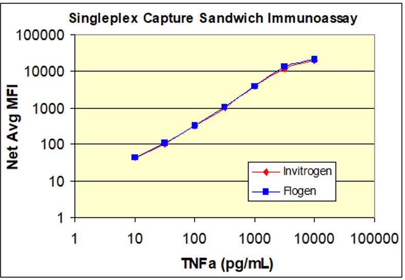
![]() Results demonstrated that Flogen SAPE exhibits a mean fluorescence intensity nearly identical to that of Invitrogen SAPE in sandwich immunoassay applications.
Results demonstrated that Flogen SAPE exhibits a mean fluorescence intensity nearly identical to that of Invitrogen SAPE in sandwich immunoassay applications. -
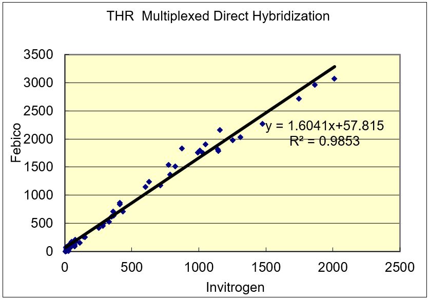
![]() Flogen SAPE was evaluated alongside Invitrogen SAPE using a DNA direct hybridization model assay. The results demonstrated a strong correlation between the two, with an R² value of 0.9853, indicating comparable performance in signal expression.
Flogen SAPE was evaluated alongside Invitrogen SAPE using a DNA direct hybridization model assay. The results demonstrated a strong correlation between the two, with an R² value of 0.9853, indicating comparable performance in signal expression.
References and Citations
- Protein Biomarkers of Cardiovascular Disease and Mortality in the Community. (n.d.). ResearchGate. Retrieved August 18, 2022, from https://www.researchgate.net/publication/326375426_Protein_Biomarkers_of_Cardiovascular_Disease_and_Mortality_in_the_Community/fulltext/5b48a3faa6fdccadaec770c6/Protein-Biomarkers-of-Cardiovascular-Disease-and-Mortality-in-the-Community.pdf
TDS/COA/MSDS
| Cat. No. | Product | TDS | COA | MSDS |
|---|---|---|---|---|
| 1SA (I, II, III, IV, V) | Streptavidin-RPE Conjugate (Type I, II, III, IV, V) |
 |
 |
 |